Medical Imaging for Pets
Medical imaging includes various techniques that allow us to take photos and images of the inside of your cat’s or dog’s body. Your vet might recommend this kind of exam for various reasons so they can take a look at your pet’s organs. At Hôpital vétérinaire des Trois-Lacs, we can perform X-rays, ultrasounds and endoscopies.
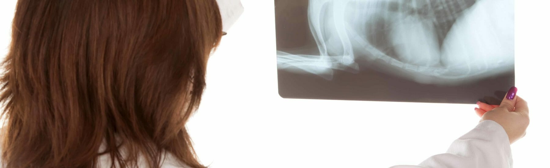
What are X-rays used for?
We use X-rays to take photos of your pet’s spine, internal organs, tissues and joints. They allow us to have a look inside your pet’s body to see if they might have:
What are ultrasounds used for?
Unlike X-rays, which take fixed images, ultrasounds use sound waves to take pictures of organs. These live images allow us to see what’s going on inside your pet’s body. Ultrasounds are often used to examine the organs of the area surrounding the abdomen, thorax and neck, including the:
What is endoscopy used for?
Endoscopy is a medical imaging technique used to examine a pet’s digestive system to look for things that can’t easily be identified with X-rays, ultrasound or even surgery.
What are the benefits of endoscopy?
The biggest benefit of endoscopy is that it’s a non-invasive way to reach places that are usually hard to get to. This reduces your pet’s pain and allows them to recover faster. It also enables us to examine very small cats and dogs.